The Foundation Model for the Neurodiverse Brain
AQAL is the world's first AI foundation model that predicts how autistic minds experience sight, sound, and language — mapping 20,484 cortical points in real time.
Understanding Neurodiversity at Scale
Understanding how autistic brains process the world has required individual brain scans — expensive, slow, and impossible for many.
AQAL changes this. A foundation model that predicts how any neurodiverse brain responds to any stimulus — transforming months of lab work into seconds of computation.

See AQAL in Action
Low sensory load. The visual cortex registers the door opening — both NT and ND brains respond similarly at this point. Minimal divergence.
AQAL Architecture
A proprietary tri-modal pipeline purpose-built for predicting neurodiverse brain activity.
Multi-Modal Encoding
Specialized encoders process sight, sound, and language independently.
Universal Integration
A deep transformer fuses modalities into brain-aligned representations.
Neurodiverse Mapping
Transforms predictions to neurodiverse patterns from real brain recordings.
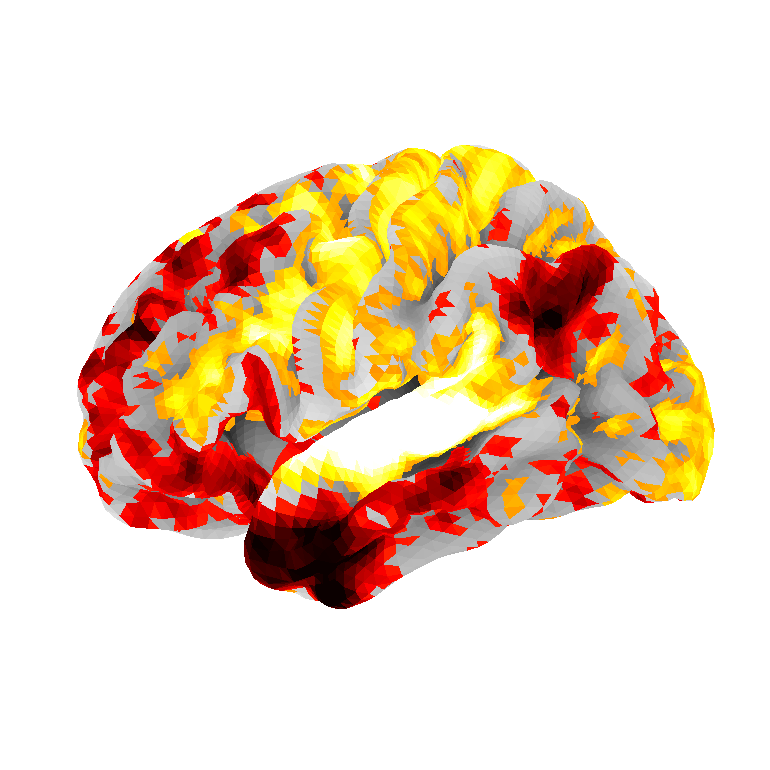
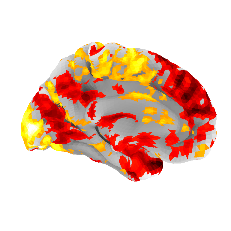
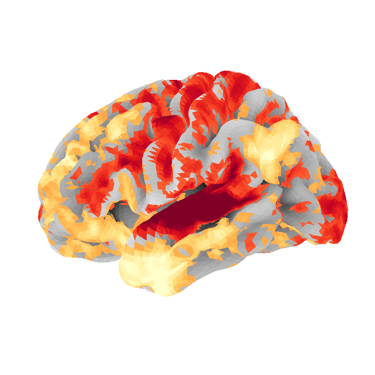
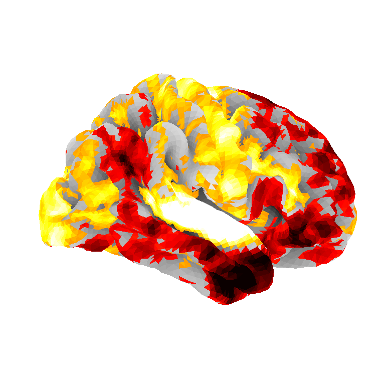
Connectivity Maps
Heatmaps showing where brain wiring differs between autistic and neurotypical individuals. Generated from 1,545 real fMRI brain scans across 36 clinical sites.
Brain Region
One of 100 parcels the brain is divided into. Each handles a specific function — vision, movement, language, emotion, etc.
Connection
A pair of brain regions that communicate with each other. With 100 regions, there are 4,950 possible pairs to test.
Connectivity
How strongly two regions are linked. Measured by how closely their activity patterns correlate over time in an fMRI scan.
t-statistic
A number measuring how different ASD and TD groups are for a given connection. Larger = bigger difference. Can be positive (ASD stronger) or negative (TD stronger).
FDR Correction
A noise filter. When you test 4,950 connections, ~248 will look significant by pure chance. FDR ensures at most 5% of your findings are false alarms.
Network
A group of brain regions that work together. The 7 major networks: Visual, Somatomotor, Dorsal Attention, Salience, Limbic, Control, and Default Mode.
1,002 Significant Connections
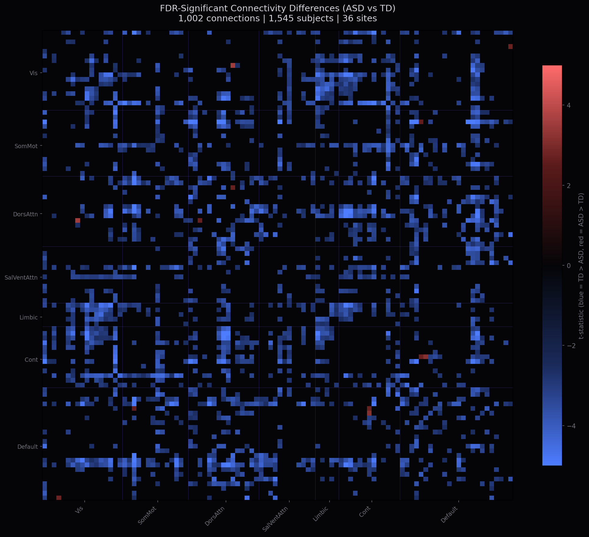
Each bright pixel represents a statistically reliable wiring difference between ASD and TD brains. Red = stronger in ASD, blue = stronger in TD. Only connections surviving FDR correction are shown.
How to read this map
Each row and column is one of 100 brain regions, grouped by network (labels on axes).
A bright red pixel at row X, column Y means that connection is significantly stronger in autistic brains.
A bright blue pixel means it's significantly stronger in neurotypical brains.
A dark/black pixel means no reliable difference was found for that connection.
The purple grid lines separate the 7 brain networks so you can see which networks are most affected.
Most Affected Brain Networks
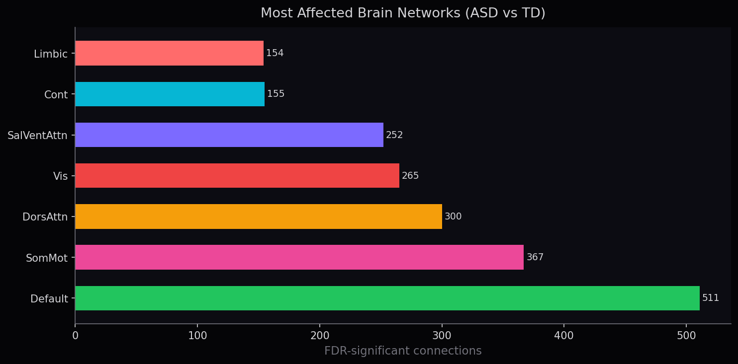
Default Mode Network shows the most affected connections (511), followed by Somatomotor (367) and Dorsal Attention (300). This aligns with known autism neuroscience — DMN alterations affect self-referential processing and social cognition.
Self-reflection, mind-wandering, social thinking. Most affected — autistic brains show altered internal processing.
Movement and body awareness. Differences here relate to motor planning, stimming, and physical coordination.
Focus and concentration. Altered wiring can explain hyperfocus on interests or difficulty shifting attention.
Processing what you see. Autistic brains often notice more detail but can be overwhelmed by busy scenes.
Filtering what matters vs background. Altered salience = difficulty ignoring the AC hum when someone is talking.
Planning and decision-making. Relates to preference for routine and difficulty with unexpected changes.
Emotion and reward. Differences here affect emotional regulation — feeling things more intensely or differently.
Full Connectivity Landscape
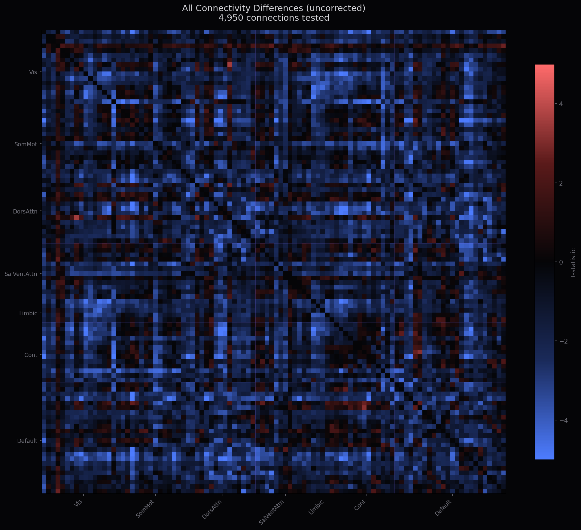
All 4,950 connections before FDR filtering. Many of these are noise — the FDR map above shows only the reliable 1,002.
AQAL Scaling Laws
AQAL follows a scaling law: accuracy increases log-linearly with more brain data. No plateau reached.
What AQAL Understands
AQAL maps how sensory processing differs across six key brain networks.
Visual
Auditory
Social
Default Mode
Salience
Motor
Powered by AQAL
The foundation model enables practical tools for autism accessibility.
Sensory Audit
LiveUpload video of any space. Get second-by-second sensory analysis, accessibility score, and actionable recommendations.
Brain Comparison
LiveSide-by-side neurotypical vs neurodiverse brain activation maps for any stimulus input.
Sensory Passport
Coming SoonPersonalized sensory profile through 5-minute calibration. Portable document for schools and clinics.
Neurotrack
In DevelopmentTherapy progress tracking with dashboards, developmental milestones, and sensory-linked reporting.
Roadmap
Foundation model trained. Statistical brain transform. Live API.
FDR-corrected. Site harmonization. Age stratification. Bootstrap uncertainty. Single consortium (871 subjects).
Dual-consortium (1,545 subjects, 36 sites). 1,002 FDR connections. Deployed.
GPU fine-tuning. Sensory subtypes. Video input. Age-specific models.
Clinically validated. 10K+ subjects. Sensory Passport. Published metrics.
Real-time processing. EEG integration. Wearable support. 100K+ subjects.
Foundation model for all neurodiversity. ADHD, SPD, anxiety. 400K+ subjects.
Build with AQAL
API access for researchers, clinics, schools, and companies building accessible products.